Development of Frog: Fertilization & its effects
Fertilization in frogs is external and aquatic. The critical events following the entry of the sperm into the egg set the stage for the entire body plan, establishing bilateral symmetry and the future dorsal-ventral axis.
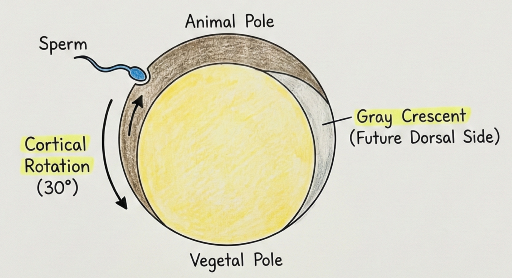
- Sperm Entry: Occurs in the animal hemisphere. Triggers completion of Meiosis II and formation of fertilization membrane (prevents polyspermy).
- Cortical Rotation: The outer cortical cytoplasm rotates ~30° towards the sperm entry point.
- Gray Crescent: This rotation exposes a lighter, pigment-free region of cytoplasm directly opposite the sperm entry site. This area is the Gray Crescent.
- Axis Formation: The Gray Crescent marks the future dorsal side of the embryo and the site of gastrulation initiation. The sperm entry side becomes the ventral side.
Cleavage
Cleavage is a series of rapid mitotic divisions of the zygote. A key feature is that there is no increase in the overall mass or size of the embryo, only an increase in cell number. The resulting cells are called blastomeres.

- Type of Cleavage: Frog cleavage is holoblastic (complete) but unequal due to the large amount of yolk in the vegetal pole.
- 1st & 2nd Cleavage: Both are meridional (vertical), passing through the animal-vegetal axis.
- 3rd Cleavage: This is latitudinal (horizontal) and occurs above the equator, closer to the animal pole.
- Result: This unequal division forms 8 cells of different sizes: 4 small micromeres at the animal pole and 4 large, yolk-laden macromeres at the vegetal pole.
- Rate: Division is much faster in the yolk-poor animal pole than in the yolk-rich vegetal pole.
Morulation and Blastulation
As cleavage continues, the embryo progresses from a solid ball of cells (morula) to a hollow ball (blastula). The formation of this cavity is essential for the subsequent movements of gastrulation.

- Morula: A transient, solid ball of approximately 16-64 blastomeres. It resembles a mulberry.
- Blastulation: As cleavage continues, the cells secrete a fluid into the center of the embryo, creating a cavity.
- Blastula: The resulting hollow sphere of cells.
- Blastocoel: The fluid-filled cavity within the blastula.
- Eccentric Position: Due to the larger size of the vegetal cells, the blastocoel is displaced towards the animal pole and is not centrally located.
Gastrulation: Initiation & Morphogenetic Movements
Gastrulation is the most dramatic stage, involving extensive morphogenetic movements of cells to rearrange the single-layered blastula into a three-layered gastrula. This process establishes the ectoderm, mesoderm, and endoderm.

- Gastrulation: The process of forming the three primary germ layers through cell migration.
- Epiboly: The prospective ectoderm cells at the animal pole divide and spread downwards to envelop the entire embryo.
- Invagination: A group of cells called bottle cells changes shape and sinks inwards on the dorsal side, creating a small groove, the blastopore.
- Involution: Cells from the marginal zone (presumptive mesoderm and endoderm) roll over the lips of the blastopore and move into the interior.
- Dorsal Lip: The upper rim of the blastopore. Its cells act as Spemann’s Organizer, a crucial signaling center.
Gastrulation: Completion & Germ Layer Formation
As cell movements continue, the three germ layers are established, the original blastocoel is obliterated, and a new cavity, the primitive gut, is formed.

- Archenteron: A new cavity formed by the invaginating endoderm, which becomes the primitive gut.
- Blastocoel Disappearance: The migrating endoderm and mesoderm cells gradually obliterate the original blastocoel.
- Yolk Plug: Large, yolky endodermal cells are encircled by the blastopore lips, forming a plug.
- Germ Layers: The embryo now has three distinct layers: outer Ectoderm, middle Mesoderm, and inner Endoderm.
- Fate of Blastopore: In frogs, the blastopore becomes the anus. The mouth forms later (deuterostome development).
Organogenesis: Neurulation (Formation of Nerve Cord)
Neurulation is the first major stage of organogenesis, where the neural tube (future CNS) is formed from the dorsal ectoderm. This process is induced by the underlying notochord.

- Induction: The notochord sends signals to the overlying dorsal ectoderm, causing it to thicken into the neural plate.
- Neural Plate & Folds: The plate’s edges rise to form neural folds, creating a central neural groove.
- Neural Tube: The folds move inwards and fuse, forming a hollow neural tube, which will become the brain and spinal cord.
- Neural Crest Cells: Cells from the fold tips migrate away to form pigment cells, ganglia, etc.
Organogenesis: Notochord and Coelom Formation
Alongside neurulation, the mesoderm differentiates into distinct regions, forming the notochord (supportive rod) and the coelom (body cavity).

- Notochord: Formed from dorsal mesoderm (chordamesoderm). It is a temporary supportive rod and signaling center.
- Mesoderm Differentiation: The mesoderm divides into Epimere (forms vertebrae, muscle), Mesomere (forms kidneys, gonads), and Hypomere.
- Coelom Formation (Schizocoely): The hypomere splits into two layers. The space between them is the coelom.
- Somatic & Splanchnic Layers: The outer somatic layer lines the body wall; the inner splanchnic layer covers the gut.
Blastula vs Gastrula Comparison
| Feature | Blastula | Gastrula |
|---|---|---|
| Cavity Present | Blastocoel | Archenteron & remnant of Blastocoel |
| Number of Layers | One (Blastoderm) | Three (Ecto-, Meso-, Endoderm) |
| Key Process | Cleavage & Cavitation | Morphogenetic Movements (Invagination, etc.) |
| Cell Fate Determined? | Partially (Animal vs. Vegetal) | Yes, fate is fixed |
MCQ Practice (Test Your Knowledge)
- The Gray Crescent, formed after fertilization, marks the future:
a) Anterior side
b) Ventral side
c) Dorsal side
d) Posterior side - Frog cleavage is best described as:
a) Holoblastic and equal
b) Meroblastic and discoidal
c) Holoblastic and unequal
d) Superficial - The cells of the dorsal lip of the blastopore, known as Spemann’s Organizer, are primarily responsible for:
a) Forming the endoderm
b) Inducing the neural tube
c) Forming the coelom
d) Becoming the epidermis - In the developing frog embryo, the blastopore eventually becomes the:
a) Mouth
b) Anus
c) Notochord
d) Neural tube - The coelom in frogs is formed by the splitting of which mesodermal layer?
a) Epimere
b) Mesomere
c) Hypomere
d) Chordamesoderm
FAQs – Development of Frog
What is the gray crescent in frog development?
It is a pigment-free region exposed after cortical rotation, opposite the sperm entry site, marking the future dorsal side.
Why is frog cleavage unequal?
Because the vegetal pole contains more yolk, slowing division there and producing smaller micromeres and larger macromeres.
What initiates gastrulation in frog?
Bottle cells invaginate on the dorsal side to form the blastopore, and involution begins at the dorsal lip.
How is the coelom formed in frog?
The hypomere splits into two layers; the space between them becomes the coelom (schizocoely).
What happens to the blastopore in frog?
In frog development, the blastopore becomes the anus; the mouth forms later.
